The Skin Barrier: A Complex Protective System
The skin barrier is not just a physical shield; it is a dynamic system composed of multiple interdependent components that work together to maintain homeostasis.
1. Physical Barrier Function
The outermost layer of the skin, the stratum corneum, acts as a fortress that prevents excessive water loss (trans-epidermal water loss, TEWL) and shields the body from external insults. This layer is composed of:
- Corneocytes – Flattened, dead keratinocytes that act like "bricks" in the skin's defense.
- Lipid Matrix – Ceramides, cholesterol, and free fatty acids form a structured “mortar” that holds the corneocytes together and maintains waterproofing.
When this structure is compromised, the skin loses moisture and becomes prone to irritation, inflammation, and secondary infections.
2. Immunological Barrier
The skin is an immunologically active organ that detects and responds to microbial and allergenic threats. It contains:
- Langerhans cells – Antigen-presenting cells that activate immune responses against pathogens.
- Keratinocytes – These produce antimicrobial peptides (AMPs) that inhibit bacterial growth.
- Cytokines and Chemokines – Signaling molecules that regulate inflammation and coordinate immune responses.
A weakened barrier can lead to an inappropriate immune response, as seen in atopic dermatitis, where allergens penetrate the skin and trigger excessive inflammation.
3. Microbial Barrier: The Role of Skin Microbiota
The skin is home to a diverse microbial community, including commensal bacteria that help protect against pathogenic overgrowth. A balanced microbiome:
- Competes with harmful microbes for nutrients and space.
- Regulates local immune responses.
- Produces antimicrobial substances to suppress pathogens.
Disruptions in this microbial ecosystem can lead to dysbiosis, predisposing pets to infections like pyoderma or Malassezia dermatitis.
Skin Barrier Defects in Atopic Dermatitis
Research shows that pets with AD have structural and biochemical abnormalities in their skin that impair its protective function. The key changes include:
Defective Lipid Composition
The skin barrier relies on intercellular lipids (ceramides, cholesterol, and fatty acids) to maintain integrity.
- Studies indicate that ceramide levels are significantly reduced in atopic dogs compared to healthy ones (Olivry et al., 2019).
- This defect increases trans-epidermal water loss (TEWL), making the skin more permeable to allergens and irritants.
Disrupted Stratum Corneum (SC) Structure
The stratum corneum, the outermost layer of the epidermis, is often thinner and more fragile in atopic dogs.
- Defective corneocyte adhesion allows allergens (dust mites, pollens, molds) to penetrate more easily, triggering inflammation.
Filaggrin and Epidermal Barrier Proteins
- Filaggrin is a key protein that helps maintain the integrity of the stratum corneum.
- Filaggrin mutations have been linked to barrier dysfunction in atopic humans and dogs (Marsella et al., 2020).
- Other structural proteins, such as involucrin and loricrin, are also reduced, weakening skin resilience.
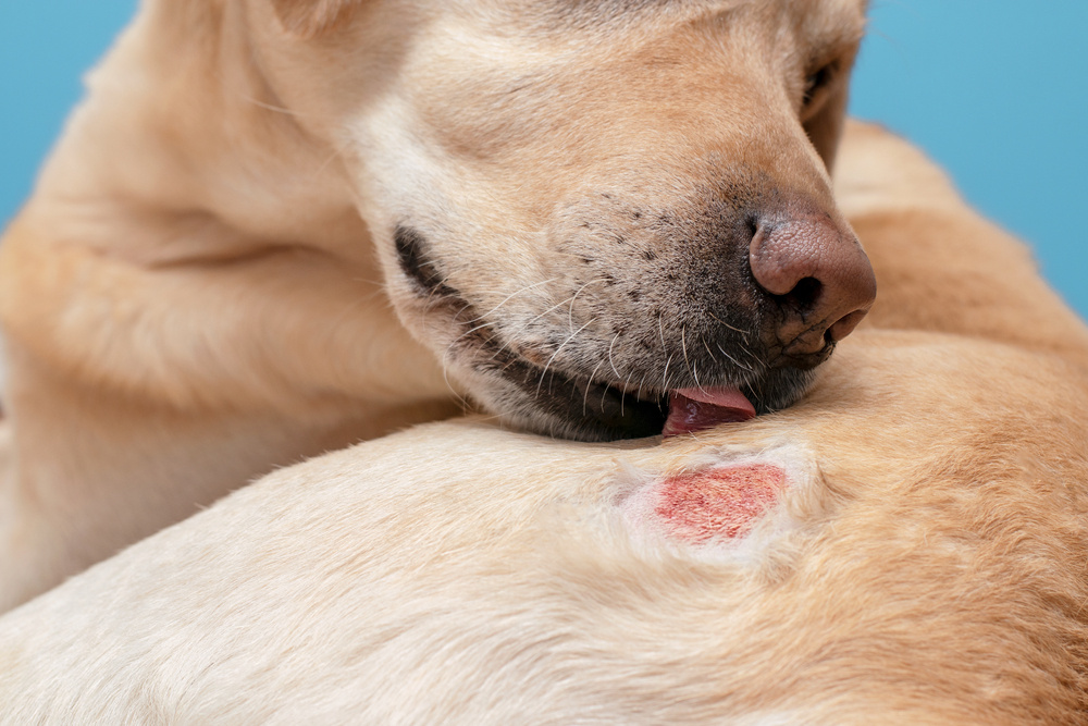
Increased Microbial Colonization (Staphylococcus & Malassezia)
- The weakened barrier allows pathogenic bacteria (e.g., Staphylococcus pseudintermedius) and yeast (Malassezia pachydermatis) to proliferate.
- Staphylococcal infections further disrupt the barrier, creating a vicious cycle of inflammation, secondary infections, and pruritus.
- Decreased antimicrobial peptide (AMP) production in atopic dogs reduces their ability to fight off infections naturally.